Hair Examination

Forensic Hair Examination
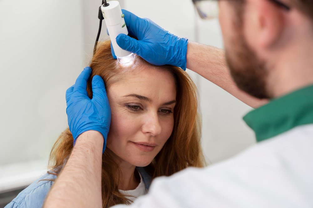
Forensic Hair Examination is one of the foundational pillars of trace evidence analysis. Hair is composed primarily of the protein keratin, making it highly resistant to chemical decomposition and environmental degradation. Because hair is shed continuously by humans and animals, it is frequently transferred during physical altercations, sexual assaults, break-ins, and vehicular accidents. The principle of Locard’s Exchange Postulate—"every contact leaves a trace"—is perfectly exemplified by the transfer of hair between a suspect, a victim, and a crime scene. A comprehensive microscopic examination of hair can provide investigators with invaluable corroborative evidence, helping to reconstruct timelines, verify testimonies, and narrow down suspect pools.
In forensic investigations, hair analysis serves a dual purpose: morphological comparison and biological assessment for DNA suitability. Morphological comparison involves examining the physical and structural characteristics of the hair under high-powered microscopes to determine its species of origin, somatic origin (the part of the body it came from), and whether it shares class characteristics with a known individual. If a hair possesses a follicular tag or root tissue, it transcends trace evidence and becomes biological evidence, highly suitable for nuclear DNA extraction. Even without a root, the hair shaft contains mitochondrial DNA (mtDNA), which can be sequenced to establish maternal lineage, a critical tool in missing persons cases and historical investigations.
Aspire Forensics provides meticulous and scientifically rigorous Forensic Hair Examination in Noida. Our state-of-the-art laboratory is equipped with advanced stereomicroscopes and comparison microscopes, allowing our experts to conduct side-by-side analyses of questioned and known hair samples. We assist law enforcement agencies, defense attorneys, private investigators, and corporate entities in solving complex cases by identifying and characterizing trace hair evidence recovered from crime scenes, clothing, weapons, and vehicles.
Clients seeking the best hair examination services in Noida depend on our strict adherence to internationally recognized forensic protocols. We ensure that every strand of hair is meticulously documented, measured, and photographed before any destructive testing is considered. This careful approach guarantees that the evidence remains viable for court presentation and potential re-examination by opposing counsel.
Furthermore, for legal teams requiring top forensic hair examination in Greater Noida, our certified examiners provide detailed, objective, and unbiased reports. We clearly articulate the scientific weight of our findings, explaining the difference between an inclusion (where a hair is consistent with a known source) and a definitive identification (which requires subsequent DNA analysis).
Scientific Principles and Hair Morphology
To understand forensic hair examination, one must understand the microscopic anatomy of a hair strand. A hair shaft is composed of three distinct anatomical layers: the cuticle, the cortex, and the medulla. The unique combination of these three layers provides the forensic examiner with a wealth of information regarding the origin and history of the hair.
The Cuticle: The outermost layer of the hair consists of overlapping, scale-like cells that point toward the tip of the hair. The pattern of these scales (coronal, spinous, or imbricate) is the primary feature used to distinguish human hair from animal hair. Humans typically possess an imbricate (flattened) scale pattern, whereas many animals exhibit intricate spinous or coronal patterns. The cuticle also protects the inner layers from chemical and environmental damage.
The Cortex: Located beneath the cuticle, the cortex is the main body of the hair shaft. It consists of spindle-shaped cortical cells aligned parallel to the length of the hair. The cortex is forensically significant because it contains pigment granules—the melanin that gives hair its color. The distribution, shape, size, and density of these pigment granules vary significantly among individuals and racial groups, making the cortex a primary area of focus during microscopic comparison. Furthermore, the cortex may contain structural features such as cortical fusi (small air spaces) and ovoid bodies, which serve as additional points of comparison.
The Medulla: The central core of the hair is called the medulla. It can be continuous, interrupted, fragmented, or entirely absent. The medullary index—the ratio of the diameter of the medulla to the diameter of the entire hair shaft—is a critical metric. In humans, the medullary index is generally less than one-third, whereas in animals, it is typically one-half or greater. The appearance of the medulla can also help identify specific animal species, such as the distinct lattice structure found in deer hair or the vacuolated structure found in dog hair.
Methodology of Forensic Hair Examination
The forensic examination of hair follows a systematic, multi-tiered approach. It begins at the crime scene or in the evidence receiving room, where trace evidence must be handled with utmost care to prevent loss or cross-contamination. Examiners use clean forceps, trace evidence vacuums, or adhesive tape lifts to recover hairs from clothing, furniture, and victims. Once secured, the evidence is transported to the laboratory under a strict chain of custody.
The first phase of laboratory analysis is macroscopic examination. The examiner observes the gross characteristics of the hair without magnification. This includes noting the color, length, form (straight, wavy, or curly), texture, and any obvious signs of damage or chemical treatment, such as bleaching or dyeing. These preliminary observations help triage the evidence and decide which hairs are most probative to the investigation.
The second phase is stereomicroscopic examination. The hair is placed under a stereomicroscope to examine its surface features in three dimensions. The examiner looks for the presence of trace materials adhering to the hair, such as blood, semen, soil, or paint. They also examine the root end (proximal end) to determine the phase of growth at the time the hair was removed. Hairs naturally shed are typically in the telogen phase and have a bulbous, club-shaped root. Hairs forcibly removed during a struggle are often in the anagen (active growth) phase and may have a follicular tag attached—a rich source of nuclear DNA.
The third and most critical phase is the comparison microscope examination. The questioned hair (recovered from the scene) and known exemplar hairs (collected from a suspect or victim) are mounted on glass slides using a specific mounting medium. They are placed on a comparison microscope, an instrument composed of two compound microscopes connected by an optical bridge. This allows the examiner to view both hairs side-by-side in the same field of view. The examiner evaluates dozens of microscopic characteristics, including pigment distribution, cuticular thickness, medullary structure, and signs of disease (such as pili annulati) or damage (like crushing from a blunt force impact).
Key Determinations in Hair Analysis
Species Determination
Analyzing scale patterns and the medullary index to definitively state whether a hair is of human or animal origin based on microscopic examination.
Somatic Origin
Determining from which part of the human body the hair originated (e.g., scalp, pubic, facial, or limb) based on diameter and cross-sectional shape.
DNA Suitability
Assessing the root structure to determine if the hair is suitable for standard STR nuclear DNA profiling or if mitochondrial DNA (mtDNA) sequencing is required.
Significance in Criminal and Civil Investigations
Hair evidence plays a pivotal role in a wide array of investigations. In cases of sexual assault, the cross-transfer of pubic hairs between the victim and the assailant can corroborate a victim's testimony, even if no other biological fluids are recovered. In hit-and-run investigations, hairs embedded in the windshield, grill, or undercarriage of a suspect vehicle can link that specific vehicle to the victim, providing a crucial piece of physical evidence that refutes a driver's alibi.
Additionally, hair acts as an historical chronological record of an individual's life. As hair grows at an average rate of one centimeter per month, chemical substances circulating in the bloodstream—such as drugs, poisons, heavy metals, and prescription medications—are incorporated into the hair shaft. While our laboratory focuses primarily on morphological and biological examination, the documentation of the hair's physical state (such as a distinct dye line indicating a recent color treatment) can establish vital timelines. If a victim was held captive, the growth of the hair root and changes in nutritional indicators within the hair can provide clues to the duration of captivity.
Exclusion is just as important as inclusion. If a questioned hair recovered from a crime scene exhibits entirely different microscopic characteristics than the suspect’s known hair, the suspect can be reliably excluded as the source of that specific hair. This exclusionary power is vital in preventing wrongful convictions and redirecting investigations toward the actual perpetrator.
Limitations of Microscopic Hair Comparison
It is vital to understand the scientific limitations of hair examination. Microscopic hair comparison is considered class evidence, not individualizing evidence. This means that while an examiner can state that a questioned hair is "microscopically consistent" with a known sample, they cannot state with absolute certainty that it came from that specific individual to the exclusion of all others in the world. Human hair exhibits natural variation, even on the same person's head.
Because of these limitations, modern forensic science uses microscopic hair examination as a screening tool. It is used to associate evidence with a suspect or to exclude a suspect entirely. If an association is made, the ultimate confirmation is achieved by passing the hair on for DNA analysis. Nuclear DNA from the root or mitochondrial DNA from the shaft provides the individualizing statistical weight required in modern courtrooms. Aspire Forensics ensures that all reports clearly state these limitations, maintaining strict scientific integrity.
Frequently Asked Questions
-
1. Can you get DNA from a hair without a root?
Traditional nuclear DNA (which identifies a specific person) requires the cellular material found in the root or follicular tag. However, the hair shaft itself contains mitochondrial DNA (mtDNA). While mtDNA cannot identify a specific individual, it can trace maternal lineage and is highly valuable for excluding suspects or identifying remains.
-
2. How many known hairs do you need for a comparison?
Because hair varies naturally on a single person's body, we require a robust exemplar sample. A standard known sample consists of at least 25 to 50 pulled hairs from various regions of the scalp (front, back, left, right, and crown) to capture the full range of natural variation.
-
3. Can you tell a person's age or gender from their hair?
Microscopic examination alone cannot determine a person's age or gender with certainty. While infant hair may appear fine and short, and graying hair indicates advanced age, these are not definitive metrics. Gender can only be definitively proven if chromosomal DNA is successfully extracted from the root.
-
4. How can you tell if a hair was forcibly removed?
When examined microscopically, a naturally shed hair will have a smooth, club-shaped root. A hair that has been pulled out with force during a struggle will typically have an elongated, stretched root, often with living tissue (the follicular tag) still attached to it.